Scanning electron microscope advantages and disadvantages in imaging components and applications.
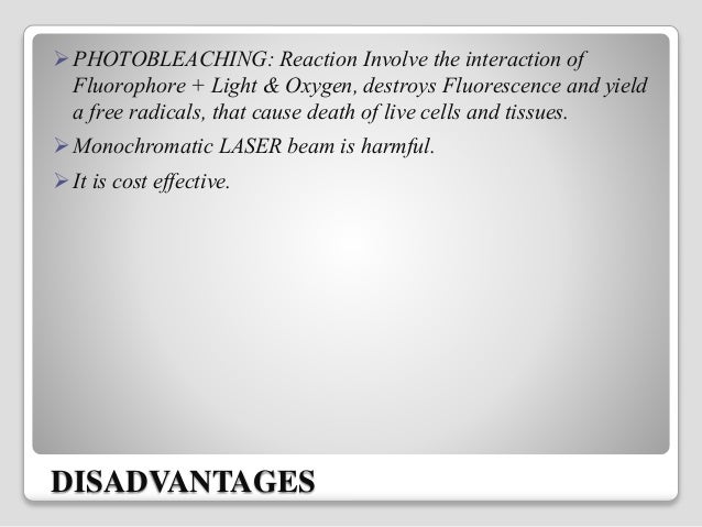
Disadvantages of laser scanning microscopes.
Advantages of confocal laser scanning microscopy industrial applications of confocal microscopy thin film profiling.
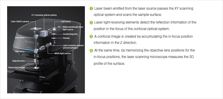
The laser scanning microscope passes a laser beam through an objective lens to illuminate a single point in an object.
Laser scanning confocal microscopy is a significant advance in the field of optical microscopy primarily because it permits sample visualization deep within living and fixed cells tissues and other samples.
A scanning electron microscope sem is a powerful magnification tool that utilizes focused beams of electrons to obtain information.
A the use of microscopy to observe and investigate different types of cell and cell structure in a range of eukaryotic organisms to include an appreciation of the images produced by a range of microscopes.
Confocal is a powerful tool but it does have some limitations.
Advantages and disadvantages of confocal microscopy.
The primary advantage of laser scanning confocal microscopy is the ability to serially produce thin 0 5 to 1 5 micrometer optical sections through fluorescent specimens that have a thickness ranging up to 50 micrometers or more.
Capturing multiple two dimensional images at different depths in a sample enables the.
Fred brakenhoff developed a scanning confocal microscope in 1979 21 while almost simultaneously colin sheppard contributed to the technique with a theory of image formation 22.
Expired practical laser scanning confocal microscope designs were translated into working instruments by several investigators.
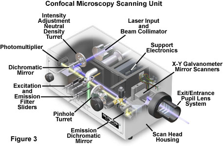
Confocal microscopy most frequently confocal laser scanning microscopy clsm or laser confocal scanning microscopy lcsm is an optical imaging technique for increasing optical resolution and contrast of a micrograph by means of using a spatial pinhole to block out of focus light in image formation.
Comparing to a wide field detection taking a snapshot of.
Speed a typical confocal uses raster scanning which means it scans the specimen point by point.
Can produce 3d.
Here are 3 quick ones.
The thickness of the coating can be determined by observing the 2 peaks in the axial intensity variation.
When investigating multilayer structures the true surface of a substrate can be observed through a surface coating.
Where electrons are used to form images.
The high resolution three dimensional images produced by sems provide topographical morphological and compositional information makes them invaluable in a.
Laser scanning confocal microscope.
It provides the ability to collect sharply defined optical sections from which three dimensional renderings can be created.